The question of pigment stability is receiving increasing attention in scientific publications on keratopigmentation. It determines the reliability of the outcome, patient safety, and the credibility of the discipline. Which biological mechanisms and formulation parameters determine how well a corneal pigment retains its properties over time?
Key Takeaways
Four factors determine the long-term stability of a keratopigmentation pigment:
the chemical composition of the formulation, which determines chemical resistance and MRI compatibility.
these influence how well the pigment remains within the implantation area.
the cellular response of the corneal stroma, particularly phagocytosis by fibroblasts.
femtosecond laser implantation parameters, particularly the incision, laser power, and tunnel depth, which directly influence pigment retention and long-term stability within the stroma.
What Is Pigment Stability in Keratopigmentation?
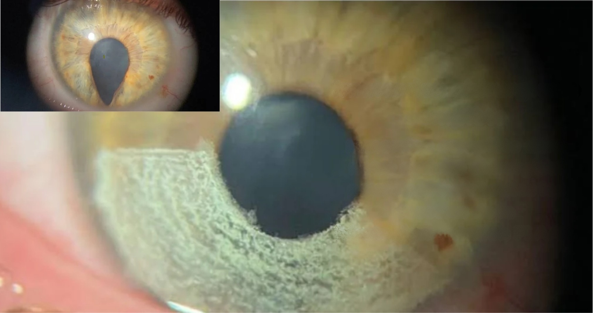
Pigment stability refers to the ability of a pigment implanted in the corneal stroma to retain its chromatic properties over time, without any change in its visual appearance. It differs from biocompatibility: a pigment that is well tolerated biologically may still undergo colorimetric changes after implantation.
The corneal stroma, an avascular tissue, provides a more stable environment than the skin. The absence of blood vessels limits rapid cellular turnover and slows the redistribution of particles. This is one reason why keratopigmentation, or corneal tattooing, can offer better pigment retention than skin tattooing under optimal conditions. However, this stability depends as much on pigment formulation as on the implantation technique and the individual biological response of the corneal tissue.
Source:
Oftalmologia Domani
.
Why Is Stability a Determining Factor in Medical Practice?
A review published in 2026 in Cornea identifies concerns regarding standardization, stability, and durability as the main barriers to the wider adoption of cosmetic keratopigmentation, highlighting the lack of long-term data on these aspects.¹ This observation reflects the concerns of many clinicians: outcome stability is what distinguishes a medically defensible technique from a procedure whose long-term results remain unpredictable.
For an implantable corneal device, longevity is an essential consideration. Patients live with the result for years, sometimes decades. An unexpected chromatic change, even a minor one, can alter the appearance of the eyes and raise clinical questions that practitioners must be able to address. This is why documented clinical follow-up remains one of the most reliable criteria for assessing pigment stability under real-world conditions.²
Which Factors Influence Pigment Retention Over Time?
The chemical composition of the pigment is the primary factor. A study published in 2026 in Experimental Eye Research establishes that mineral pigments used in keratopigmentation are chemically stable and that any color change necessarily involves pigment migration away from its original implantation area, rather than chemical degradation.³ This finding directs attention toward biological mechanisms as well as formulation chemistry.
Controlling particle size distribution is therefore an essential formulation requirement that directly influences pigment behavior within the corneal tissue.
The tissue’s cellular response represents a third factor. The same 2026 study identifies phagocytosis by corneal fibroblasts as one of the biological mechanisms contributing to pigment loss: stromal cells can engulf pigment particles and cause their redistribution.³ This phenomenon is aggravated by an inflammatory response, which may itself be promoted by certain compositions or by disruption of the basement membrane during implantation.
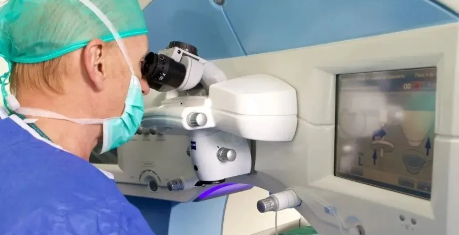
The implantation technique directly influences long-term stability. Intrastromal placement using a femtosecond laser embeds the pigment within the thickness of the stroma, where it is protected by the overlying corneal layers. Professor Alió documented this as early as 2016: this technique reduces the risk of pigment loss compared with superficial approaches while preserving the integrity of the ocular surface.⁶
Prof. Jorge Alió — Source.
What Role Does Formulation Play in Pigment Stability?
Formulation is the parameter over which the manufacturer has the greatest direct control, starting from the pigment design stage.
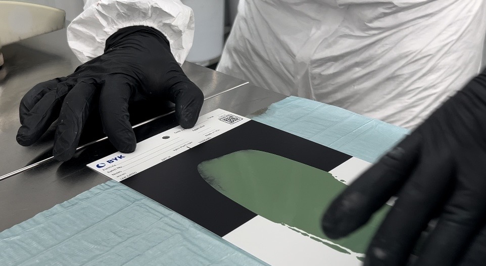
Laboratoires BIOTIC Phocea, certified to EN ISO 13485, manufacture their medical pigments using high-purity raw materials consisting of mineral and organic pigments selected for their chromatic stability. Each batch undergoes physical quality controls covering color, density, viscosity, and flow time, as well as spectrophotometric measurements and assessment in a light booth with controlled color temperature. Sterilization is performed by gamma irradiation at a dose exceeding 25 kGy, with batch-number traceability, ensuring sterility for five years. No preservatives are added, and the dispersing agent is plant-derived.⁷
The issue of iron oxides illustrates the impact of chemical composition on long-term safety. Available data indicate that pigments containing iron oxides can generate MRI artifacts and, under the influence of the magnetic field, migrate within the corneal tissue.⁴ A study specifically conducted on BIOTIC Phocea formulations containing iron oxides, published in Cornea in July 2026 by Dr. Movshovich et al., presents more nuanced findings: no heating or pigment movement was observed, but local distortion of the corneal MRI signal was detected.⁵ These findings highlight the need to evaluate each formulation individually rather than drawing conclusions based on pigment categories.
It is precisely because each formulation must be assessed individually that BIOTIC Phocea chose to reformulate certain raw materials rather than wait for broader data to become available: a black pigment based on activated carbon, free from iron oxides and nanoparticles, and a white pigment based on boron nitride, formulated without titanium dioxide. European and US patent applications for these innovations were announced at the Kolor Congress in Dubai, following more than two years of R&D.⁸
How Do Pigments Behave Under Different Lighting Conditions, and Why Does This Determine How Natural the Result Looks?
The natural iris does not have a uniform, fixed color. Under bright light, its surface structures become more visible and its color appears more intense. In dim lighting, the perceived color becomes softer. A keratopigmentation pigment produces a more natural-looking result when it reproduces these optical variations rather than creating a flat, static color.
Aesthetic keratopigmentation emerged directly from reconstructive practice: it was through observing the quality of results obtained when correcting damaged irises, their uniform coverage, and their natural appearance under different lighting conditions that the technique was extended to purely aesthetic indications.⁶ This origin continues to shape formulation requirements today: the BIOCHROMAEYES® and BIOCHROMAEYES® Aesthetic shades were developed to match natural iris colors, with respective palettes of 12 shades for BIOCHROMAEYES® and 16 shades for BIOCHROMAEYES® Aesthetic designed for this purpose.
Pigment chemistry directly influences this optical behavior. According to clinical observations collected from practitioners, composite formulations combining several complementary mineral shades produce light scattering that more closely resembles that of a natural iris than pure monochromatic shades, particularly under varying lighting conditions. These same observations indicate that very light shades may fade slightly over several years in cases of significant light exposure, reinforcing the value of mineral formulations for their resistance to photodegradation.
Conclusion
The stability of a keratopigmentation pigment is measured by its ability to maintain its chromatic properties within living tissue over the long term. Research published in recent years has documented this issue with increasing precision, identifying the biological mechanisms involved and the formulation parameters that address them. In a discipline where outcomes are assessed at six months, one year, and sometimes beyond, pigment stability is inseparable from clinical quality.
Frequently Asked Questions
What Is Pigment Fading in Keratopigmentation?
Are Keratopigmentation Pigments Compatible with MRI?
How Can the Quality of a Keratopigmentation Pigment Be Assessed?
Why Does the Visual Appearance of a Pigment Change Depending on the Light?
Notes and References
¹ Alió JL, El Bahrawy M, et al. Cosmetic Keratopigmentation Works and Can Be Used as a New Aesthetic Solution to Change the Apparent Color of the Eye. Cornea. 2026. [PubMed link to be added]
² Alió JL, et al. Therapeutic Keratopigmentation Vis-à-Vis in Blind Eyes: Complications and Outcomes in a Series of 184 Eyes. Frontiers in Medicine. 2025. DOI: 10.3389/fmed.2025.1700788.
³ Cellular and Cytokine Responses of Corneal Fibroblasts Exposed to Keratopigmentation Inks In Vitro. Experimental Eye Research. 2026. [PubMed link to be added]
⁴ Magnetic Resonance Imaging and Keratopigmentation: A Systematic Review and Ex Vivo Study. Cornea. 2026. [PubMed link to be added]
⁵ Movshovich A, et al. MRI Safety of Iron Oxide–Containing Pigments in Keratopigmentation. Cornea. Published July 31, 2026. DOI: 10.1097/ICO.0000000000004199.
⁶ Alió JL, Rodriguez AE, El Bahrawy M, et al. Keratopigmentation to Change the Apparent Color of the Human Eye: A Novel Indication for Corneal Tattooing. Cornea. 2016. [PubMed link to be added]
⁷ Manufacturing data, certifications, and quality processes: Laboratoires BIOTIC Phocea. Our Medical Pigments — EN ISO 13485 Certificate.
⁸ Characteristics of the new BIOCHROMAEYES® Aesthetic formulations: activated-carbon-based black pigment, boron-nitride-based white pigment, and European and US patent applications. Innovation & Safety — BIOTIC Phocea.
European patent application EP25173297.0: COMPOSITION FOR KERATOPIGMENTATION WITH VEGETABLE CARBON BLACK AND WITHOUT NANO OR IRON BLACK.
European patent application EP25167552.6: COMPOSITION FOR KERATOPIGMENTATION WITHOUT TITANIUM DIOXIDE.